ATTENTION: The works hosted here are being migrated to a new repository that will consolidate resources, improve discoverability, and better show UTA's research impact on the global community. We will update authors as the migration progresses. Please see MavMatrix for more information.
Show simple item record
| dc.contributor.author | Nandwana, Vikas | |
| dc.contributor.author | Kang, Shishou | |
| dc.contributor.author | Shi, Shifan | |
| dc.contributor.author | Jia, Zhiyong | |
| dc.contributor.author | Thompson, G. R. | |
| dc.contributor.author | Nikles, David E. | |
| dc.contributor.author | Harrell, J. W. | |
| dc.contributor.author | Poudyal, Narayan | |
| dc.contributor.author | Liu, J. Ping | |
| dc.date.accessioned | 2010-10-13T20:41:57Z | |
| dc.date.available | 2010-10-13T20:41:57Z | |
| dc.date.issued | 2009 | |
| dc.identifier.citation | Published in Journal of Applied Physics 101 | en_US |
| dc.identifier.issn | 0021-8979 | |
| dc.identifier.uri | http://hdl.handle.net/10106/5082 | |
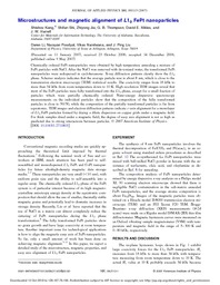
| dc.description.abstract | Chemically ordered FePt nanoparticles were obtained by high temperature annealing a mixture of FePt particles with NaCl. After the NaCl was removed with de-ionized water, the transformed FePt nanoparticles were redispersed in cyclohexanone. X-ray diffraction patterns clearly show the L10 phase. Scherrer analysis indicates that the average particle size is about 8 nm, which is close to the transmission electron microscopy (TEM) statistical results. The coercivity ranges from 16 kOe to more than 34 kOe from room temperature down to 10 K. High resolution TEM images reveal that most of the FePt particles were fully transformed into the L10 phase, except for a small fraction of particles which were partially chemically ordered. Nano-energy dispersive spectroscopy measurements on the individual particles show that the composition of the fully transformed particles is close to 50/50, while the composition of the partially transformed particles is far from equiatomic. TEM images and electron diffraction patterns indicate c-axis alignment for a monolayer of L10 FePt particles formed by drying a dilute dispersion on copper grids under a magnetic field. For thick samples dried under a magnetic field, the degree of easy axis alignment is not as high as predicted due to strong interactions between particles. | en_US |
| dc.description.sponsorship | Department of Physics, University of Texas at Arlington. Center for Materials for Information Technology, The University of Alabama | en_US |
| dc.language.iso | en_US | en_US |
| dc.publisher | AIP | en_US |
| dc.subject | Iron alloys | en_US |
| dc.subject | Platinum alloys | en_US |
| dc.subject | Nanoparticles | en_US |
| dc.subject | Annealing | en_US |
| dc.subject | X-ray diffraction | en_US |
| dc.subject | Magnetic particles | en_US |
| dc.subject | Particle size | en_US |
| dc.subject | Transmission electron microscopy | en_US |
| dc.subject | Magnetic structure | en_US |
| dc.subject | Coercive force | en_US |
| dc.subject | Solid-state phase transformations | en_US |
| dc.title | Microstructures and magnetic alignment of L10 FePt nanoparticles | en_US |
| dc.type | Article | en_US |
| dc.identifier.externalLink | http://www.uta.edu/ra/real/editprofile.php?onlyview=1&pid=1866 | en_US |
| dc.identifier.externalLinkDescription | Link to Research Profiles | en_US |
Files in this item
- Name:
- mic_and_mag.pdf
- Size:
- 287.0Kb
- Format:
- PDF
This item appears in the following Collection(s)
Show simple item record